This post is a collaborative effort by Becca (Blindodiaries) and Erin (Whistlererin). We had a lot of fun putting it together.
To our fellow Blindos out there, how many times have you tried to explain that you’re VI by simply saying “I’m visually impaired.” or “I have a sight problem.” Then, the sighted person you are explaining this to either says, “Oh, so you’re blind,” or “But you can see, right?” Quite a few times? Yeah us too. So today, we’re going to do our best to explain and show some the different degrees of vision, and how the world looks to a person with low vision or legal blindness. Every VI person sees differently, and even these examples show only a small sample of the different ways people see, or the different methods used to cope with a visual impairment.
Major misconceptions about vision:
If you’re blind you only see black
If you’re visually impaired that means you’re blind.
If you see the tree across the street that means there is no possible way you can’t read the words right in front of your face.
If you can see the words right in front of your face then you MUST be able to see the details on the tree 5-10ft in front of you.
You can transplant an eye (Note: The only part of the eye that can currently be transplanted is the cornea.)
If you appear to be looking at something, you can see all of it clearly.
If you’re carrying a white cane but looking at your watch, you must be faking!
If only it was that simple! But vision is much more complex than that. Your eye itself is the most complex organ in your body. At the bottom of the post, we have included a list of the parts of the eye for reference, in case you’re not sure what we’re talking about when we refer to the retina or cornea. We have also included some of the most common ways of measuring visual acuity.
So here, we’ve put together some stories of VI people, including ourselves. Each story is based on a lot of research and real people that we know, although we have changed the names and some of the identifying details to protect privacy.
The most obvious beginning is with our own vision! First, me, Becca: imagine having 20/400 vision (see Snellen Eye Chart below, if you’re unsure what this means). This means that that E is non existent! If I remove my contact lens and look at that chart I don’t even see the chart I see a blurry white square shaped thing. I have 20/400 vision uncorrected.

It makes it a bit tricky to find curbs, recognize faces or read signs. To be honest, the majority of my blurry issues are due to not having a lens in the back of my eye because of my glaucoma. When it comes to my ROP which has caused my blood vessels to grow in abnormal ways and at one point caused me to be cross eyed my issues are different. I mostly have issues with depth perception and focusing. Because my Nystagmus (my eyes/head shake when I am trying to focus on an object) makes it difficult to focus I have a tendency to squint a lot. This doesn’t mean I can’t see it it just means it is a bit of a pain (figuratively and literally) to do so.
~~~~~~~~~~~~~~~~~~~~~~~~~~~~~~~~~~~~~~~~~~~~~~~~~~~~~~~~~~~~~
For me, Erin, Having severe myopia (nearsightedness), migraines, and some CVI (cortical visual impairment) are what cause problems for me. I already wrote an in-depth post on it, so if you want the whole enchilada, read here. In a nutshell, if you take the picture of my living room, decorated for Christmas last year:

And see it how I see it, you get lens distortion and blurred-out periphery. Throw in eyestrain, dry eyes and some other issues, and you sort of get the picture.
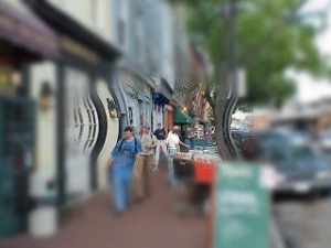
We can also take the street scene from Becca’s example, and you’ll see how hard it is to find things, particularly curbs or small children directly in front of my feet.
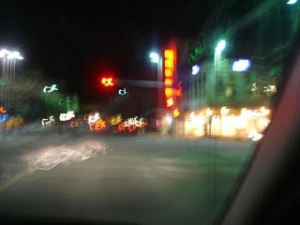
With a migraine, I prefer not to wear my glasses, but to wear dark sunglasses, as I become quite light-sensitive. So a street scene grows blurry and dark, even in the daylight.
Enough about us! Here are some other examples of vision loss.
~~~~~~~~~~~~~~~~~~~~~~~~~~~~~~~~~~~~~~~~~~~~~~~~~~~~~~
Retinitis Pigmentosa
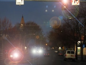
A person who has Retinits Pigmentosa, known commonly as RP, which is a degenerative eye condition reports a gradual (over many years) loss of peripheral vision, usually from the outside in. Because the fovea, or central part of the retina, sees details the best, people with RP can read books or a computer screen, but have trouble walking around or finding something he/she set down.
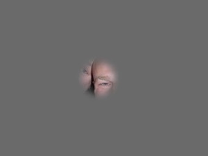
Because RP causes night-blindness, someone with RP has trouble recognizing faces in a dim restaurant, or getting around at night. In spite of the progressive vision loss, most people keep an upbeat attitude. The progressive nature of RP, however, they find difficult to deal with sometimes, as they wake up to new losses or constantly have to adjust how they cope.
~~~~~~~~~~~~~~~~~~~~~~~~~~~~~~~~~~~~~~~~~~~~~~~~~~~~~~
Glaucoma
Glaucoma is a condition in which pressure in the eye causes damage to the optic nerve, resulting in blind spots or loss of peripheral vision.
At times, the brain will fill in the blind spots with something that it thinks ought to be there, causing either hallucinations, or a scene in which the user does not realize is incorrect. Left untreated, these can multiply until all vision is lost.

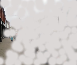
My (Erin’s) daughter, Abi, who is three, has Congenital Glaucoma, meaning she was born with this condition. Although she cannot tell us definitively what she sees, she seems to see only a little bit out of the extreme left side of her vision, as shown in the photo above.When she walks around, she can see large objects just to her left, and when looking at a toy, she holds it close to her face on the left side. Mostly, though, she uses her hearing and touch to navigate the world. Because she has always seen this way, she is not bothered in the least by her vision, and to her the world looks perfectly normal.
~~~~~~~~~~~~~~~~~~~~~~~~~~~~~~~~~~~~~~~~~~~~~~~~~~~~~~
Macular Degeneration
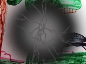
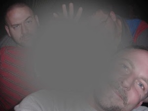
Macular Degeneration (AMD) is commonly found in adults 65 and older. This disease causes you to lose your central vision. The majority of my (Becca’s) retired clients have this disease. Though they lose their central vision, which is the part of the eye you focus with, they are still able to live normal lives. The major issues that come with this eye disease include have trouble with reading, trouble with colors and losing objects with a slight turn of the head. Interestingly enough the black spot in the picture below is actually pretty accurate when ever a client describes their vision they always say “there is a block spot in front of my eye.”
This child’s drawing might be handed to a grandparent to admire.
With AMD, it’s difficult to see the detail in the picture, as the central vision alone is responsible for seeing fine details.
~~~~~~~~~~~~~~~~~~~~~~~~~~~~~~~~~~~~~~~~~~~~~~~~~~~~~~
Cataracts
This eye disease can come at any age. It consists of a white film covering the lens in your eye. Some times this is visible to the naked eye and other times it is not. Cataracts can be removed with surgery when they get a mature stage. Having a cataract will cause everything you see to become blurry as well as foggy. This will also cause issues with light sensitivity, I (Becca) have been told by clients that things seemed washed out and very dark even on a bright day.
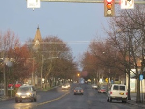
Normal view of a street
Another condition that might result in such a view is corneal scarring, which obscures a view, and causes light sensitivity and loss of acuity.
~~~~~~~~~~~~~~~~~~~~~~~~~~~~~~~~~~~~~~~~~~~~~~~~~~~~~~
Hyperopia
As people age, close object become blurred; however, this can usually be corrected with glasses.
~~~~~~~~~~~~~~~~~~~~~~~~~~~~~~~~~~~~~~~~~~~~~~~~~~~~~~
Color Blindness
There are several types of color blindness. A person with normal color vision sees the scene below:
A commonly-held myth is that all color-blind people see the world in black and white. While there are a few who report seeing no color at all, most people do see some colors.
The most common type of color blindness is red/green color blindness, in which those two colors cannot be distinguished from one another.

Some conditions which result in both color loss and loss of acuity make it much harder to distinguish objects.
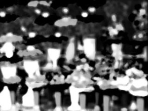
Other impairments can result in less color perception.
~~~~~~~~~~~~~~~~~~~~~~~~
Photophobia
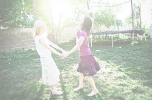
Photophobia is an extreme sensitivity to light. Many conditions have this. If a person with typical vision sees a scene like this:
A visually impaired person can see a painful, washed-out scene instead.
~~~~~~~~~~~~~~~~~~~~~~~~~~~~~~~~~~~~~~~~~~~~~~~~~~~~~~
Achromatopsia
This rare genetic condition affects the cones in the retinas, and results in partial or total color blindness, extreme light sensitivity and loss of acuity. In the daylight, where a sighted person sees this:

Someone with Achromatopsia sees a scene something like this:

Since someone who has Achromatopsia has had a stable visual condition her whole life, she may rarely think about her vision at all. She might enjoys an active lifestyle; in fact I (Erin) read a bio of an athlete in the Iditarod Sled-dog race who has this condition. To cope with the light sensitivity, people wear prescription red-tinted sunglasses or contact lenses, which block out the color spectrum that causes he most discomfort to her.
~~~~~~~~~~~~~~~~~~~~~~~~~~~~~~~~~~~~~~~~~~~~~~~~~~~~~~
Stargardt’s Disease
Like Age-related Macular Degeneration, Stargardt’s results in loss of central vision, although a few people I (Erin) have spoken with say that their peripheral vision has been lost as well. This condition is somewhat rare also.
~~~~~~~~~~~~~~~~~~~~~~~~~~~~~~~~~~~~~~~~~~~~~~~~~~~~~~
Motion Blindness
Along with other visual problems, some people report being unable to track moving objects, such as a ball moving.
Depth or Convergence Problems
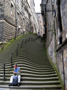
Many conditions that cause a visual impairment affect each eye differently. They also may cause distortion in the visual field. If an unaltered staircase looks like this:
Someone with a visual impairment may see the staircase as looking distorted like this:
Or they may have difficulty focusing on just one image or gaining enough clarity to make out the details in individual steps.
Leber’s Congential Amaurosis (LCA)
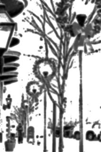
We are told that LCA is like looking through the “snow” that used to show up on TV screens when there was no signal. It usually gets bad enough that there isn’t useable vision after a while, although it varies from person to person.
So, there you have it! From our own research and talking to friends, clients, online acquaintances, doctors and others, we’ve given you a little glimpse into the interesting world of the visually impaired. A legally blind person rarely sees nothing but black, and some visual tasks are certainly more difficult than others, depending what he or she sees. We hope this brings a little more knowledge and understanding, as well as respect for those who navigate their world in a different way.
******************************************************
For reference:
The human eye is about 2.5 cm in length and weighs about 7 grams. Light passes through the cornea, pupil and lens before hitting the retina. The iris is a muscle that controls the size of the pupil and therefore, the amount of light that enters the eye. Also, the color of your eyes is determined by the iris.
The vitreous or vitreous humor is a clear gel that provides constant pressure to maintain the shape of the eye. The retina is the area of the eye that contains the receptors (rods and cones) that respond to light. The receptors respond to light by generating electrical impulses that travel out of the eye through the optic nerve to the brain.
Parts of the Eye
· Aqueous Humor: Clear, watery fluid found in the anterior chamber of the eye.
· Choroid: Layer of blood vessels that nourish the eye; also, because of the high melanocytes content, the choroid acts as a light-absorbing layer.
· Cornea: Transparent tissue covering the front of the eye. Does not have any blood vessels; does have nerves.
· Iris: Circular band of muscles that controls the size of the pupil. The pigmentation of the iris gives “color” to the eye. Blue eyes have the least amount of pigment; brown eyes have the most.
· Lens: Transparent tissue that bends light passing through the eye. To focus light, the lens can change shape by bending.
· Pupil: Hole in the center of the eye where light passes through.
· Retina: Layer of tissue on the back portion of the eye that contains cells responsive to light (photoreceptors).
· Rods: Photoreceptors responsive in low light conditions.
· Cones: Photoreceptors responsive to color and in bright conditions.
· Sclera: Protect coating around the posterior five-sixths of the eyeball.
· Vitreous Humor: Clear, jelly-like fluid found in the back portion of the eye. Maintains shape of the eye.
If any of the aforementioned parts of the eye are damaged then your vision will become worse. This does not automatically mean that you will completely lose your vision but you will have various kinds of issues.
Visual Acuity (VA): The clarity of sharpness of vision. For example, “If you have 20/20 vision, you can see clearly at 20 feet what should normally be seen at that distance. If you have 20/100 vision, it means that you must be as close as 20 feet to see what a person with normal vision can see at 100 feet.” (www.aoa.org)
Currently a Snellen Chart is the most common test used to asses VA’s. “A standard eye chart is necessary to make comparisons and to record people’s visual acuity. The most common chart used in most doctors’ offices is the Snellen eye chart. In 1862, a Dutch Ophthalmologist, Dr. Hermann Snellen, devised this eye chart. He determined that there was a relationship between the sizes of certain letters viewed at certain distances. A copy of the Snellen chart may be found here.
The Snellen eye chart has a series of letters or letters and numbers, with the largest at the top. As the person being tested reads down the chart, the letters gradually become smaller. Many other versions of this chart are used for people who cannot read the alphabet. The Tumbling E chart has the capital letter “E” facing in different directions and the person being tested must determine which direction the “E” is pointing, up, down, left, or right. A Broken Wheel vision test is one that can be used for children or those who cannot read the alphabet and the person being tested must tell which card has the broken wheels on the pictured car. Another type of eye chart that can be used is a picture chart with common pictures of different sizes.”(http://www.mdsupport.org/library/acuity.html)
The Snellen Eye chart breaks your vision up into fragments of 20/10, 20/15, 20/20,20/25, 20/30, 20/40.20/50,20/100,20/200. And yes that big E at the top of the chart is the 20/200 line.










Very interesting article! I was researching articles for a blog post for Braille Month, but I am definitely going to link to your informative and, pardon the pun, eye-opening article here. Thank you!